
Rotator Cuff Disorders
Information published by IRCOS about Rotator Cuff Disorders: relevant practitioners, available content and related documents to help you prepare for your consultation.
The bones of The head of the humerus is located at the top of the arm: it is round, covered with cartilage and articulates with the glenoid, a rather flat joint area of the scapula (or scapula) which is the bone which connects the upper limb to the thorax. The scapula continues posteriorly with a bony prominence which returns above the tendons of the rotator cuff: the acromion.
THE ROTATOR CAP
The rotator cuff takes its name from the appearance of the tendons that cover the head of the humerus: they cover it like hair covers a head, hence the term “cuff”
The rotator cuff tendons come from four muscles whose fibers are attached to the scapula: the muscle fibers transform into tendons as they approach the humeral head.
There are four muscles (and therefore four tendons):
1°) The supraspinatus muscle (or supraspinatus or supraspinatus)
– It takes its name from its position at the level of the scapula: it is above a bony element called the spine of the scapula
– It is the muscle whose tendon is almost always the first c – It gives the shoulder its power, for example its ability to lift heavy objects.
2°) The infraspinatus muscle (or infraspinatus or infraspinatus)
– It takes its name from its position at the level of the shoulder blade: it is below the spine
– Its tendon is often affected secondly, after the supraspinatus, in tendinitis and ruptures, but to a lesser extent.
– It is the main muscle responsible for the external rotation movement of the shoulder: ability to direct the hand outwards when the elbow is glued to the body or ability to lift the hand upwards when the arm is horizontal
3°) The subscapularis muscle (or subscapularis)
– It takes its name from the area of the shoulder blade where its fibers attach: the subscapular fossa.
– Its tendon is more rarely affected in its entirety than that of the infraspinatus, however it is frequently affected in its upper part.
– It is partially responsible (along with other muscles) for the internal rotation movement of the shoulder, including the ability to place the hand behind the back.
4°) The teres minor muscle (or teres minor)
– It takes its name from its shape because it is small and round!
– It is exceptionally affected by tendonitis and ruptures,
only in very evolved and very old forms, and only once the tendon of the infraspinatus muscle has completely disappeared.
Aside: the tendon of the long head of the biceps:
– The biceps, a well-known arm muscle, has two tendons in its proximal part: one is located outside the shoulder joint, the other is located in the shoulder joint and passes under the subscapularis tendons in front of the arm, then between the supraspinatus and subscapularis tendons to attach to the scapula.
– It is not considered as a rotator cuff tendon because its mechanical role in the shoulder is not very important.
– On the other hand, it is often affected by disease, because as soon as it is exposed to inflammation or rupture of the cuff tendons, and in particular the supraspinatus and subscapularis tendons, it can itself become inflammatory and painful on its own account, or even rupture as well.
THE DELTOID MUSCLE
Directly under the skin, it covers the rotator cuff.
It is the main driving muscle of the shoulder.
It is never affected in rotator cuff pathology (except following overly aggressive open surgery), which explains why the shoulder can move despite a complete tear of the rotator cuff: only the deltoid then allows part of the movement, without however providing the total power of the shoulder.
Symptomatology
PAIN
This is the master symptom.
It is always present at one point or another during the course of the disease.
It can appear suddenly, after effort or trauma, even mild. It can also occur gradually.
It is often nocturnal, sometimes it even prevents you from sleeping due to its intensity or wakes you up in the middle of the night around 4 a.m., which reflects its inflammatory nature.
During the day, it can be important but also only become annoying during certain gestures or during exercise.
It can also be present for a given period then disappear for several months or even several years before reappearing during an effort, a sudden movement, sometimes spontaneously.
At the start of the disease, it is easily calmed by simple medications such as analgesics or anti-inflammatories. It may end up resisting any treatment.
It can be linked:
– Has simple tendinitis.
– Has a tendon rupture.
Its location is very variable and there is no direct correlation between its position and tendon damage.
It is often located “in the shoulder” without precise location, changing position with time or movement.
It often radiates towards the arm, or even towards the forearm and the fingers, which can cause rotator cuff damage to be confused with an attack of neurological origin such as Cervico-Brachial Neuralgia. It often also radiates towards the neck and back, following the path of the trapezius muscle which inserts from the top of the neck to the middle of the back.
LOSS OF STRENGTH
This is the first sign we generally notice, apart from pain. It reflects the damage to the supraspinatus and is all the more important as the musculotendinous damage is significant and long-standing.
It is sometimes isolated, that is to say the only sign present, the pain having been calmed by medical treatment.
Most of the time it reflects a tendon rupture and no longer simple tendonitis.
LOSS OF MOBILITY
It is the inability to use the arm for certain gestures:
– Either requiring force (affects the supraspinatus)
– Or requiring an external rotation movement (involvement of the infraspinatus and teres minor)
– Or requiring an internal rotation movement (damage to the subscapularis)
– Either requiring a combination of these movements (damage to several tendons)
This loss of mobility has a double meaning:
Either it is present at the start of a tendon rupture, and reflects the intensity of the initial inflammation. It is then often recoverable by medical treatment, in particular by the rehabilitation component of medical treatment.
Either it is present late, after a more or less long course of the disease. It then resists medical treatment and often reflects an advanced and complete form of tendon rupture.
OTHER SYMPTOMS
– Cracking, pseudo-blockages are rarer.
– Aside: the Popeye sign: it is the appearance of a lump in the arm, often after an intense painful phase. It reflects the rupture of the tendon of the long head of the biceps.
Clinical Examination
Your surgeon will first begin with a questioning to clarify your symptoms, their intensity, their date of appearance, their evolution, their importance, the discomfort caused in daily life, work and leisure, the various treatments used until now.
The actual clinical examination allows us to assess:
– Muscle morphology.
– Active and passive mobility of the shoulder.
– Painful areas.
– the strength and resistance of the various rotator cuff tendons.
At the end of the clinical examination, your surgeon will most often prescribe additional radiological examinations, or will examine those prescribed by your attending physician.
No biological examination is useful in rotator cuff pathology.
Additional Examinations
SHOULDER RADIOGRAPHS
They are essential because they allow us to highlight
– The morphology of the acromion.
– The existence of osteoarthritis.
However, they are often normal.
ULTRASOUND OF THE ROTATOR CUFFER
It requires an operator who is highly trained in rotator cuff pathology.
It is difficult to interpret by the surgeon
It is rarely sufficiently precise to allow a therapeutic decision.
MRI (Nuclear Magnetic Resonance Imaging)
Often a good screening test, it allows a diagnosis that is sometimes sufficiently precise for the therapeutic decision.
Its interpretation is sometimes difficult:
* In small or partial tendon damage.
* In the evaluation of the extension of lesions.
The use of injected contrast product (arthroMRI) allows greater precision and provides images close to arthrography in quality.
THE ARTHROSCANNER
This is the reference exam.
It allows both:
* Accurate diagnosis, including partial or small lesions.
* Easy assessment of the extent and severity of lesions: tendon retraction, fatty degeneration, muscle atrophy.
It is almost systematically requested by the surgeon preoperatively and sometimes postoperatively to check the quality of tendon healing.
Cuff tendinitis
It is an inflammatory condition of the tendon, most often the supraspinatus, WITHOUT rupture.
It rarely occurs before the age of 40
It often affects the dominant limb and is more common in upper limb strength workers or in those who use their shoulder in repetitive movements beyond the horizontal.
It results in pain, rarely in a limitation of mobility or a loss of strength.
Its treatment is primarily medical (see treatment below). It is rarely surgical.
Cuff rupture
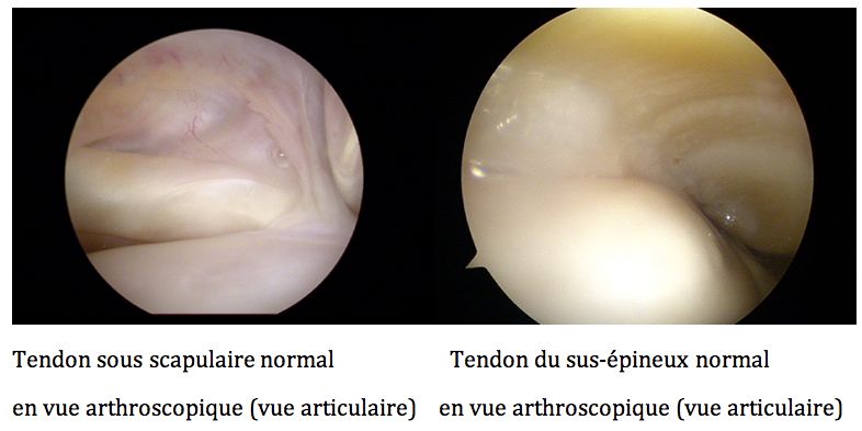
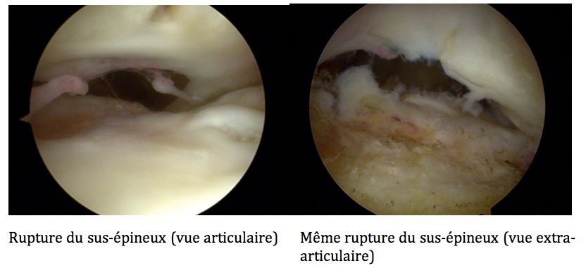
It mainly concerns the supraspinatus, often after a more or less long phase of tendinitis.
It occurs especially after the age of 50 and is then part of rotator cuff tendon disease (prolonged tendinitis then rupture of a degenerative tendon)
Exceptionally, it can be post-traumatic before the age of 50, the tendon then being healthy, but simply torn from its bony insertion by a very violent opposing movement (fall during a high-speed accident, countered movement of the shoulder, etc.)
The pain is still present, but may be reduced, in particular by prior medical treatment of the tendinitis. It can also be much more intense than with tendinitis: anything is possible at this level.
The loss of strength is constant and this is a very good sign to differentiate between simple tendinitis and a rupture: it is however necessary for your surgeon to clarify this point because it is not always easy to make a diagnosis yourself!
The loss of mobility varies depending on the old or recent nature of the rupture, the effectiveness of the treatments already carried out, and the extent of the damage.
The progression of the supraspinatus rupture always becomes worse:
– At the level of the supraspinatus itself with retraction of the tendon, muscular atrophy and fatty degeneration of the muscle.
– Towards achieving
* Either of the infraspinatus, which then worsens in the same direction as the supraspinatus, with tendon retraction, atrophy and muscular fatty degeneration: these are the so-called “postero-superior” ruptures: often a little less painful and therefore detected at an already advanced stage, that of loss of mobility for example.
* Either of the subscapularis, with the same evolution: tendon retraction, atrophy and fatty degeneration. These are the so-called “antero-superior” ruptures: often more painful and therefore detected at an earlier stage.
– After some time (from a few months to a few years), the development can lead to damage to the 3 tendons (supraspinatus, infraspinatus and subscapularis). The humeral head is then no longer held by any tendon and gradually rises to come into conflict with the acromion. The cartilage can then wear out and we witness the appearance of osteoarthritis specific to the development of rotator cuff tears: “eccentric oarthrosis” (osteoarthritis of the shoulder with rise of the head of the humerus)
Medical treatment
It should always be tried as first intention, especially in tendinitis.
It includes:
– Medications: analgesics and non-steroidal anti-inflammatories. (oral corticosteroids should be avoided as much as possible)
– Rehabilitation:
* Learning certain gestures to avoid overloading the rotator cuff tendons.
* Strengthening of adjacent muscles (deltoid, back muscles) * Adjuvant therapies: ultrasound, use of cold (cryotherapy)
* Certain actions should be avoided: pulley therapy type work or even muscle strengthening work, without direct control of the physiotherapist.
– Infiltrations:
* They use a combination of local anesthetics and locally injectable depot corticosteroids.
* They should ideally be carried out under radiological control in order to be sure to put the product in the right place: between the tendons of the rotator cuff and the acromion: so-called “subacromial” infiltration
* They are reserved for failure of first-line treatment (medications and rehabilitation)
* They must be limited to 3 per year maximum.
* It is preferable to use them in the absence of tendon rupture, therefore in cases of tendonitis without rupture, or when the tendon rupture can no longer be repaired: it is therefore useful to have tendon imaging before offering them.
Surgical treatment
It is reserved for failures of medical treatment but can be chosen immediately in certain circumstances
– Traumatic rotator cuff tear before age 50.
– Rupture of the slightly retracted rotator cuff, with good quality muscles, in an active patient with strength or repetitive work of the upper limb, generally before the age of 65.
ANESTHESIA
During the pre-anesthesia consultation which must legally take place (except in an emergency) at least 48 hours before the operation, the anesthesiologist will specify the procedures for shoulder anesthesia.
Anesthesia is usually mixed:
– Loco-regional anesthesia: by placing, under local anesthesia, a catheter around the nerves of the upper limb, at the base of the neck. This catheter is left in place for 24 to 48 hours after the operation and helps reduce immediate post-operative pain.
– general anesthesia: short and often without complete intubation, thanks to the effectiveness of the catheter above, it remains essential given *the sometimes long duration of surgical procedures,
*often uncomfortable position during the procedure,
*the need for absolute immobility of the shoulder during the surgeon's technical actions.
ARTHROSCOPIC ACROMIOPLASTY
Consists of planing the lower surface of the acromion in contact with the rotator cuff.
It is carried out
– Either for the treatment of tendinitis, in the event of failure of medical treatment, when the shape of the acromion, pointed and developed downwards, can explain the origin of the tendinitis.
– Either during the repair of the rotator cuff, to allow the repaired tendon not to be in conflict with the acromion during the healing phase which is always accompanied by a transient thickening of the repaired tendon.
It is now recommended that this acromioplasty be performed arthroscopically (see below)
Hospitalization is short: 24 to 48 hours, sometimes on an outpatient basis.
In acromioplasty without tendon repair, immobilization of the upper limb is done using a sling, removable for washing, dressing and rehabilitation, and is short-lived: 8 to 10 days.
Post-operative rehabilitation is essential.
The work stoppage is 45 days to 3 months.
Recovery of a normal shoulder takes place over a period of 3 to 6 months.
TENOTOMY OR TENODESIS OF THE LONG PORTION OF THE BICEPS UNDER ARTHROSCOPY
It consists of cutting the biceps tendon, under arthroscopy at the level of the shoulder joint (tenotomy) and possibly fixing it lower (tenodesis).
It is very often performed during rotator cuff tendon repair.
Isolated, it is rarely used and is only intended for two particular cases:
– Isolated biceps tendonitis.
– Irreparable rupture of the rotator cuff tendons with significant biceps tendinitis, in patients over 65 years old, without shoulder osteoarthritis.
ARTHROSCOPY REPAIR OF THE ROTATOR CUFFER
Arthroscopy has revolutionized shoulder surgery.
It allows a much more complete exploration of the shoulder as well as a certain number of gestures impossible to perform in the open.
It significantly decreases:
* Length of hospitalization: rarely more than 48 hours.
* Muscle damage to the deltoid: non-existent during arthroscopy.
* Certain complications: risk of infection or much less risk of hematoma.
Finally, but certainly not the most important: the aesthetics of scars are naturally significantly superior to open surgery.
Within our association, all cuff repairs have been carried out arthroscopically for 10 years now.
IN PRACTICE:
A certain number of incisions 0.5 to 1.5 cm long are made around the shoulder.
A rigid optical fiber connected to a video camera is introduced through one of these incisions in the shoulder.
Instruments allowing remote manipulation are introduced through the other incisions.
The repair of the lesions is thus carried out under direct video control by the surgeon.
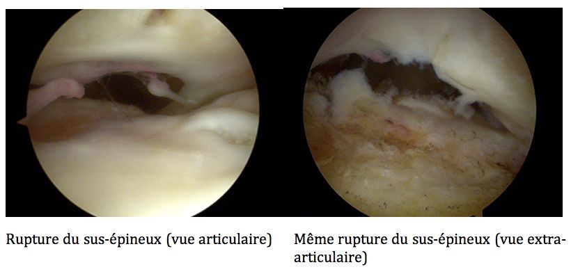
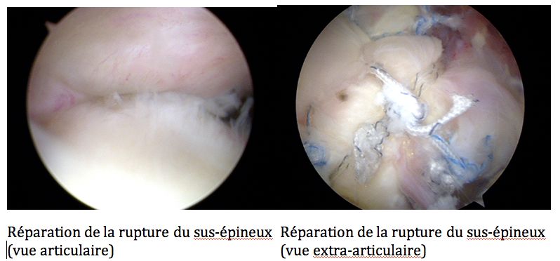
The scars are then closed with a simple stitch or simple small sticky strips (steristrip)
The shoulder is immobilized for 4 to 6 weeks depending on the action performed by a removable sling for dressing, washing and rehabilitation during this period.
At the end of this period of immobilization, the first post-operative consultation generally takes place. It allows you to:
– Proceed with the definitive removal of the immobilization sling
– Check for the absence of immediate complications. (see below)
– Check the quality of the first actions carried out during rehabilitation.
The work stoppage varies depending on the profession exercised and whether or not it is necessary during professional activity to exert effort on the repaired tendons: it varies between 3 and 6 months on average.
Recovery of a mobile and painless shoulder is effective after 3 to 6 months on average. It is often necessary to wait a year to recover normal strength, similar to the strength of the shoulder before the tendon rupture.
A check of tendon healing by CT arter is generally requested at the 6th month in order to check the quality of this healing.
TENDON TRANSFER
It is a surgery that only concerns a few patients: in fact the conditions are as follows:
– Irreparable rupture of the rotator cuff tendons, with retracted tendons and atrophic muscles.
– Patient under 65 years old.
– Failure of medical treatment.
– Preserved mobility of the shoulder (beyond 90°)
It consists of transferring in place of the retracted tendons one or more other tendons close to those of the rotator cuff.
It is a surgery that is done partially open (removal of the tendon), the transfer and attachment of the tendons in place of the rotator cuff tendons can be done arthroscopically.
We distinguish:
– Transfer of the pectoralis major muscle tendon: for irreparable ruptures of the subscapularis.
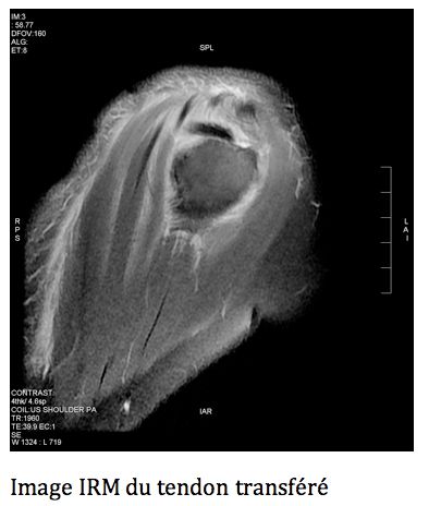
– Transfer of the latissimus dorsi muscle tendon: for irreparable ruptures of the supraspinatus and infraspinatus
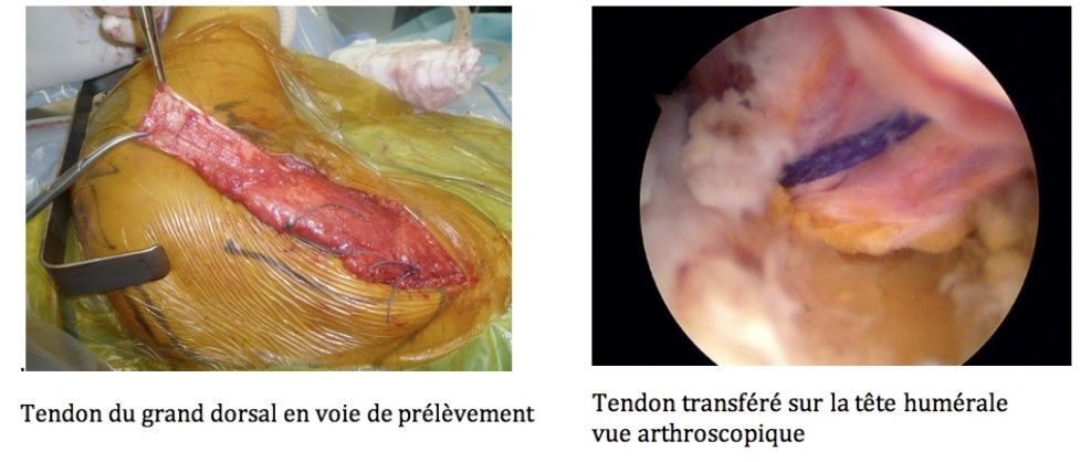
Tendon transfer surgery gives satisfactory results in reducing pain and restoring mobility but does not allow recovery of strength as good as in tendon repair.
THE REVERSE PROSTHESIS
Used in cases of eccentric osteoarthritis (shoulder osteoarthritis with ascension of the head of the humerus)
It is only offered over the age of 65 in the event of irreparable rupture of the rotator cuff tendons associated with significant osteoarthritis.
In certain exceptional cases, it can be offered in the absence of osteoarthritis, over the age of 65, in the event of irreparable rupture of the rotator cuff tendons and significant functional impotence of the shoulder (movements < 90°)
Complications
Despite advances in arthroscopy, rotator cuff surgery is not free of complications.
These are uncommon and most heal without after-effects and without the need for further surgical intervention.
However, some are more serious and may require one or more new surgical procedures to achieve a cure.
1°) Recurrence of tendon rupture
This is the main complication in terms of percentage.
It is all the more common when:
– The condition of the tendons is initially poor: retracted tendons, atrophied muscles.
– The tendons are too thin and difficult to move under traction during the procedure.
– Immobilization is not respected.
Recurrence is thus rare (<10%) for a relatively recent rupture, not very retracted, where the repair was carried out on a thick and easily movable tendon.
It can go up to 40, or even 50%, in the event of a rupture operated too late, with a retracted tendon, thin and difficult to move and atrophied muscles.
A recurrence of tendon rupture does not always give the same symptoms as before the intervention, particularly in terms of pain and sometimes only a partial loss of strength is noted. A new intervention is then not necessary.
Only the reappearance of pain, not calmed by medical treatment, can lead to a new surgical procedure:
– Or a new attempt at repair if the condition of the tendon and muscles on arthroscopy allows it.
– Or other surgical therapies: tendon transfers, reverse prosthesis.
2°) Adhesive capsulitis
It is the second most common complication: it occurs in 3 to 6% of cases.
An unpredictable complication independent of the condition of the tendon, the type of surgery, or the surgeon, it is directly linked to an “excessive” reaction of the shoulder to the surgical procedure.
It extends the recovery time, often multiplying it by 2 or 3, with recovery sometimes requiring 18 months or more.
It has no direct impact on tendon healing.
The after-effects are very rare, sometimes like partial stiffness of the shoulder in large movements, more rarely like residual pain.
The treatment is based on the fight against pain and gentle rehabilitation
3°) Infection
Exceptional in arthroscopic surgery, it is – Superficial: in 0.1 to 0.3% of cases, requiring only treatment with short antibiotic therapy and local care.
– Deep: in less than 0.1% of cases, requiring further surgical intervention and prolonged antibiotic therapy. The final result then depends on the possibility of keeping the repair equipment or not and therefore on the speed of reaction to this complication.
The clinical signs being the same in both cases: fever, inflammatory appearance of the scars, abnormal discharge, swelling of the shoulder, only your surgeon will be able to differentiate between superficial and deep infection.
4°) Much more exceptional:
– Nervous complications: partial and transient paralysis of certain muscles of the hand or upper limb, rare (0.15% of cases), most often linked to loco-regional anesthesia.
– Vascular complications: extremely rare, most often linked to anesthesia.