Meniscus
Information published by IRCOS about Meniscus: relevant practitioners, available content and related documents to help you prepare for your consultation.
Menisci
Meniscus
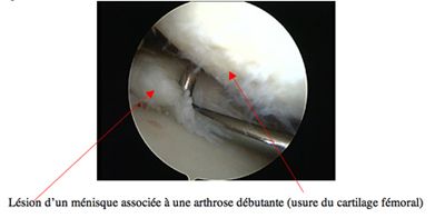
This lesion is the most common traumatic injury of the knee…
Clinical signs
You have :
– Pain on the inside or outside of the knee
– Clicking in the knee or crunching sensations
– Regular knee locks
– A recurrent effusion of the knee…
* You may have: a MENISQUE injury
*This lesion is the most common traumatic knee injury: it affects 4/10,000 women and 9/10,000 men each year.
Anatomy
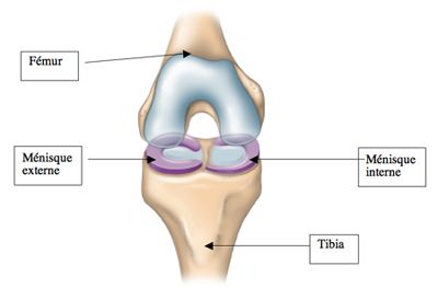
This is the knee shock absorber.
There are 2 menisci in the knee: an internal one and an external one. They are crescent-shaped: the external meniscus is a little more closed than the internal, almost 0-shaped.
They are attached to each other and to the cruciate ligaments of the knee with which they work in harmony.
They follow the movements of the femur and tibia during flexion and extension of the knee to best adapt to all situations. The internal meniscus is attached to the internal lateral ligament of the knee over its entire length: it is therefore more stable and less mobile than the external meniscus but at the same time very vulnerable to shearing movements created by rotation of the knee.
The external meniscus is not attached to the joint capsule over its entire length and is separated from the external lateral ligament: it is therefore more mobile and less stable than the internal meniscus.
Imaging
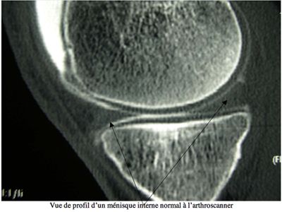
The menisci are not calcified like bone: they are therefore invisible on simple x-rays.
Two exams can visualize them:
– One via a contrast product injected into the knee: THE ARTHROSCANNER. CT arthrography is also very useful for studying cartilage.
Your doctor or surgeon will prescribe one or other of these tests depending on the type of lesion associated with the meniscal lesion they are looking for (cartilage lesion or ligament lesion)
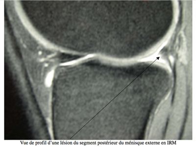
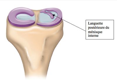
Meniscal lesions vary greatly in their shape and location:
These may be cracks, more or less mobile tabs, large displaced fragments (also called bucket handles, often responsible for acute blockages of the knee).
Cracks can be vertical, horizontal, radial.
The internal meniscus is mainly affected at its posterior part, with progressive extension towards the middle part, more rarely towards the anterior part.
The external meniscus is mainly affected at its middle part, with possible extension forwards, but also backwards.
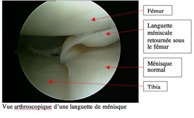
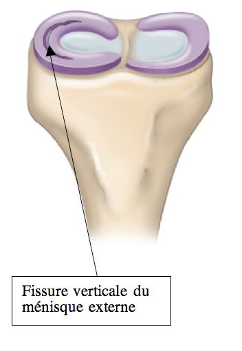
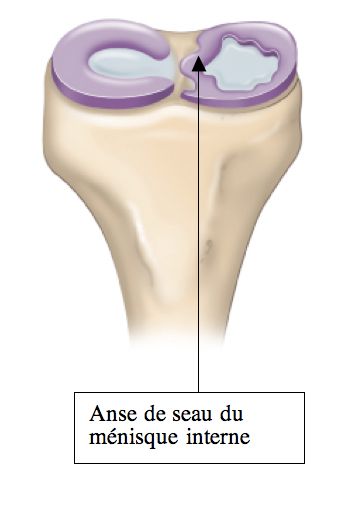
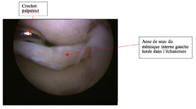
imagery
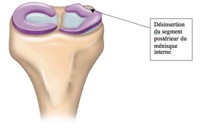
Some meniscus injuries are rarer:
– Peripheral meniscal disinsertion, often associated with an injury to the anterior cruciate ligament, it corresponds to the detachment of the meniscus from its peripheral ligamentous attachments.
– Rare lesions:
• Congenital discoid meniscus (disk-shaped meniscus, especially visible on the external meniscus in children)
• Meniscal cyst: especially external, it is always associated with a horizontal meniscal fissure. It sometimes results in a small lump visible under the skin.
• Meniscal ossicle: small ossification inside the meniscus. Images of cyst, ossicle.
Why operate
– Because the lesion is responsible for the pain and its treatment leads to healing in more than 80% of cases.
– Because the presence of a meniscal lesion can be responsible for other lesions, themselves irreversible: in particular cartilaginous lesions by abnormal friction of detached parts of the menisci on the adjacent cartilage.
The intervention
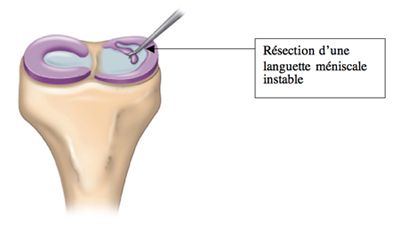
ARTHROSCOPY is the rule for meniscal surgery.
It consists of introducing a camera into the knee through a small incision and one (or more) instruments through one (or more) other incisions.
These instruments are most often used to remove the detached meniscus fragment responsible for the pain. Meniscal surgery is therefore CONSERVATIVE: it removes the responsible fragment and preserves the rest of the meniscus.
Sometimes, in the event of peripheral disinsertion, meniscal repair is possible using a system of small anchors and wires.
The anesthesia is most often loco-regional, consisting of putting the affected knee or both legs to sleep (spinal anesthesia).
The procedure is carried out AMBULATORY: the patient returns the morning of the procedure and leaves the same evening with a simple dressing, without crutches or splints, with authorization to walk immediately.
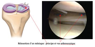
The surgical aftermath
Walking is authorized immediately, but you must know how to rest for a few days after the procedure.
Returning to work is often possible quickly, sometimes after a few days for patients who do not have to drive a vehicle and who have a sedentary job.
For patients who have a heavier job or who must drive a vehicle, a rest of 2 to 3 weeks is recommended.
A few rehabilitation sessions, started a few days after the operation, allow optimal recovery of mobility and tone of the knee.
Recovery after meniscal surgery to remove the internal meniscus usually occurs within one to two months, with the return to sports being able to begin at 2 months.
The delay is often a little longer for the external meniscus, 2 to 3 months. In the case of meniscal repair, the time is also longer, around 2 to 4 months.
Long-term results are good, even better if there is no associated lesion (ligamentous or cartilaginous)
– 90% of knees having undergone a partial internal meniscectomy (removal of a fragment of the meniscus) and 70% of external meniscectomies are perfectly painless and normal 15 years later when the meniscus lesion is isolated and of traumatic origin (no associated ligamentous or cartilaginous lesion)
– 70% of meniscal repairs result in meniscal healing.
Complications
They are rare or even exceptional (<0.1%)
Blood effusion, Infection, Algodystrophy, Phlebitis, ligament injury, vascular injury, equipment breakdown, rapid chondrolysis.
The rare complications of meniscal surgery should not be confused with the consequences of lesions sometimes associated with meniscal lesions.
1°) Thus, an injury to the ANTERIOR CRUISES LIGAMENT is responsible for INSTABILITY, the injury to the meniscus is a CONSEQUENCE of the absence of an anterior cruciate ligament. It reflects poor tolerance of instability and is a turning point towards worsening in the history of instability due to ligament rupture. Ligament reconstruction is therefore at best effective BEFORE the appearance of meniscal lesions
2°) The association with CARTILAGE lesions (or chondral lesions) is also possible: these cartilaginous lesions evolve FOR THEIR OWN ACCOUNT, hence the importance of performing meniscal surgery BEFORE their appearance. The persistence of pain, duller and present with prolonged walking, is often linked to these cartilage lesions