Ankle Ligament Pathology
Information published by IRCOS about Ankle Ligament Pathology: relevant practitioners, available content and related documents to help you prepare for your consultation.
Sprained ankle
This is the most common traumatic pathology. It affects one in 10,000 people per day. It represents up to 50% of accidents among footballers.
9 times out of ten, it concerns the external lateral ligament of the ankle.
RECENT SPRAIN
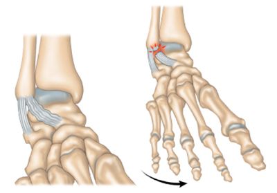
Usually, the patient “twists” the ankle. the foot going inwards.
The initial pain is acute. You can hear a crack. However, in the heat of the moment, the patient often continues his sporting activity.
The pain reappears regularly a few hours later and intensifies. The ankle swells and we sometimes notice the famous “pigeon egg” which is in fact a pocket of blood on the lateral edge of the ankle.
The lesion is variable. It may be a simple tendon “stretch”, a partial tear or a complete tearing of the external lateral ligament. Most often, it is the anterior bundle of the ligament that is injured.
Initial treatment requires “icing” the ankle in order to limit swelling and therefore pain. The ankle must be elevated. Support and movement should be limited. The prescription of analgesics and anti-inflammatories and the use of canes are useful in the initial phase.
As soon as support resumes, a flexible orthosis must be put in place, generally for 5 to 6 weeks (duration of ligament healing). This system includes 2 plastic valves connected by Velcro which prevent the ankle from twisting and recreating the injury mechanism which caused the sprain. There are many models (Airlock, aircast, axmed, etc.) which advantageously replace plaster boots, allow total support and generally do not require anticoagulants.
Around the 10th day, we will begin rehabilitation which must meet several objectives:
– fight against edema and pain, with drainage massages and ultrasound
– recovery of joint range of motion
– proprioceptive rehabilitation: this involves teaching the patient to lock their ankle, by re-teaching them to stabilize themselves on unstable platforms or balls. These exercises are essential to avoid the recurrence of the sprain and the appearance of an unstable ankle.
ankle instability
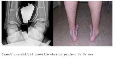
This is the recurrence of regular ankle sprains without apparent triggering reasons. The patient “twists” the ankle easily. There are several episodes per year and sports practice is frequently interrupted.
Sometimes, it is the persistence of ankle pain that leads to the diagnosis.
This will be confirmed by taking dynamic images which require twisting the ankle and confirm excessive yawning of the joint between the tibia and the talus.
Anterior drawer maneuvers are also performed where the ankle is pushed forward in relation to the leg.
The x-rays are taken in a comparative manner.
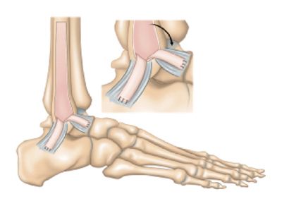
In the absence of sufficient improvement through well-conducted proprioceptive physiotherapy, it is necessary to “reconstruct” an external plan.
Most often we carry out a re-tensioning of the external plane which has healed too loosely. Depending on the quality of the remaining tendon, reinforcement is sometimes combined with the tissue lining the fibula. We then speak of a periosteal flap.
In practice, hospitalization is outpatient
The patient is immobilized by a boot for 21 days without support. Secondarily, support is authorized and rehabilitation begins. Immobilization is limited to wearing a removable orthosis for another 3 weeks.
The work stoppage is one month for a sedentary worker and 2 months for a forced worker.
Anterior ankle impingement
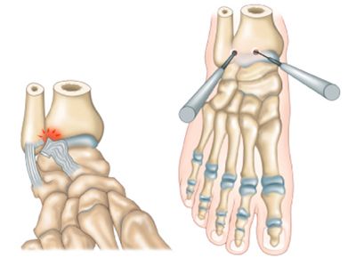
The patient mainly complains of pain on the anterolateral edge of the ankle. This discomfort predominates in a squatting position and dorsiflexion of the ankle. This is often thick and sometimes swells.
There may be a subjective feeling of ankle instability. However, dynamic images do not find objective instability. This is the consequence of repeated sprains.
Healing tissue appears after each LLE sprain. This bulky tissue can crowd the joint and get stuck in the joint during certain movements.
This is a diagnosis of elimination after carrying out a CT arthrography eliminating another cause. This examination can highlight this hypertrophic scar tissue.
A local anesthetic infiltration is carried out during the examination and relieves the patient.
Treatment consists of arthroscopic resection of the scar tissue. This procedure takes place under regional anesthesia.