
Patellar Disorders
Information published by IRCOS about Patellar Disorders: relevant practitioners, available content and related documents to help you prepare for your consultation.
What are you suffering from?
You have :
– Pain in your knee when going down the stairs.
– In a prolonged sitting position, you feel the need to extend your legs
– You have the feeling that your knee is temporarily locked
– You do not feel confident in your support
You may have a problem with your kneecap. We then speak of a pathology of the patellofemoral joint (patella = kneecap).
Anatomy
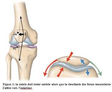
The patella is located in front of the knee. It slides at the anterior part of the femur in a cartilaginous groove called the trochlea. It is connected to the femur by the quadriceps tendon which is the union of 4 muscles. This is the most powerful muscle group in the body.
The patella is connected to the tibia by the patellar tendon.
When the knee flexes, the patella descends along the trochlea.
Centering the kneecap is essential so that the movement occurs harmoniously. The operation can be compared to that of a pulley. If the rope is poorly centered in the groove of the pulley, the rope will heat up and wear out prematurely.
It is often said that the patella joint is the “barometer” of the knee.
Any knee problem can result in patellar pain or patellofemoral syndrome.
Imaging
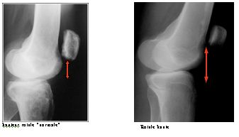
Examination of the knee by the doctor and taking simple x-rays usually make it possible to make the diagnosis.
This is a profile x-ray of the knee at 30° of flexion which allows us to assess the position of the patella in relation to the trochlea and to assess the length of the patellar tendon.
In fact, the longer it is, the more likely the kneecap is to engage poorly in the groove. We then speak of high patella.
The other important x-ray is taking a photo (called patellofemoral) allowing you to see the patella from above. This allows us to appreciate the shape of the patella and its centering in the femoral groove, the depth of which can be appreciated. If the banks of the trochlea are not raised enough, the patella can escape from its rail and become dislocated. We then speak of patellofemoral dysplasia.

Imaging
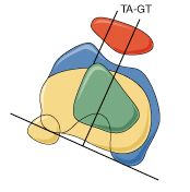
As part of a pre-operative assessment, a CT scan is sometimes requested. This examination makes it possible to measure the eccentricity of the knee extensor system.
This measure, called TAGT, is necessary before performing surgical “recentering” of the patella. We also study the tilt of the patella which reflects the primacy of one muscle group over another and which hinders engagement.
An arthroscopy is sometimes carried out with the injection of a contrast product into the joint which makes it possible to identify areas of cartilage wear.
At the end of the consultation we can distinguish 2 distinct tables:
– The unstable patella with episodes of dislocation or potentially unstable. This is a painful kneecap with radiological criteria of instability, but having never “dislocated”.
– Patellar pain syndrome or RDS. This is pain of patellar origin without any evidence of the slightest abnormality within the joint. This is a problem with the functioning of the knee, without it having the slightest architectural anomaly. The vast majority of patellar syndromes are RDS. This is an extremely common pathology among adolescents and young adults where it represents around 20% of consultations.
Rehabilitation
Rehabilitation is the main treatment for all patellar pain.
It is carried out in parallel with the reduction of physical activities and the possible taking of painkillers and anti-inflammatory drugs.
It must meet 2 objectives:
– Le premier objectif est de lutter contre la rétraction musculaire qui majore l’hyperpression sur la rotule .
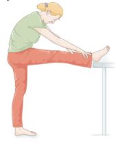
– The second is to optimize the position of the patella in relation to the trochlea to ensure the most ergonomic patellar stroke possible. It requires stretching the structures that pull the kneecap toward the outside of the leg. This is associated with strengthening the internal bundle of the quadriceps. This rehabilitation must be carried out with the knee discreetly flexed and must absolutely be carried out without weight on the tip of the foot. In fact, this presses the patella against the femur and risks increasing cartilage damage.
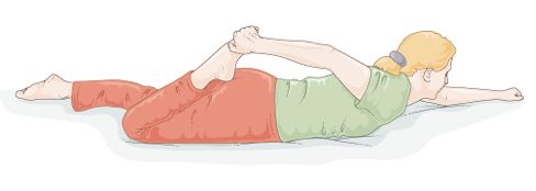
In practice, after warming up, we begin with self-stretching Knee in extension, we perform a stretching of the posterior structures.
Lying on your stomach, you try to place your heel on your buttock; stretching posterior structures.
Patellar recentering surgery
It is primarily aimed at unstable kneecaps with a history of dislocation. It will be offered after failure of rehabilitation.
The intervention generally combines the performance of 3 actions:
– First: the section of the external fin fibrous structure attracting the patella towards the outside of the knee
– Deuxièmement : le transfert de la tubérosité tibiale antérieure (TTA ) . Il s’agit du relief osseux sur lequel est inséré le tendon rotulien . Le déplacement de la TTA vers le versant interne de 10 mm permet de recentrer la. On y associe parfois un abaissement lorsque la rotule est trop haute .
– Third: plasty of the vastus medialis. We modify the angle of insertion of the quadriceps on the upper part of the patella. This gesture makes it possible to increase the attraction of the patella internally and to fight against its tilt
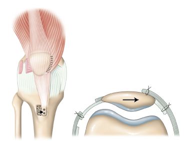
The procedure requires a vertical incision in the anterior part of the knee and the installation generally of 2 screws to fix the bone fragment.
Hospitalization is approximately 2 days.
The tube to prevent knee hematoma is removed on the 2nd day.
Full support is authorized under the cover of a removable splint to be kept for 45 days. Rehabilitation began immediately. A 2-month work stoppage is generally necessary.
Although the results are generally good, there may be reasons for dissatisfaction:
-Early: The occurrence of a hematoma sometimes requires reoperation. The occurrence of an infection is rare (less than 0.5%). The weakening of the tibia and the TTA sometimes requires careful re-education of the patient. Knee stiffness is prevented by appropriate rehabilitation.
– Secondarily: a recurrent dislocation of the patella has already been described, especially since the knee carries several architectural defects. Rarely, some patients complain of persistent patellar pain.
Arthroscopic treatment of patellar pathologies
Arthroscopy allows more limited procedures to be performed than conventional surgery. It is used to make isolated external fin sections.
In the event of cartilage damage to the patella, it is sometimes used to clean the joint.
This surgery takes place on an outpatient basis.